
Photons create golden boxes, electrons lead to shells
16 September 2026
EurekAlert!: [https://www.eurekalert.org/news-releases/1144375]
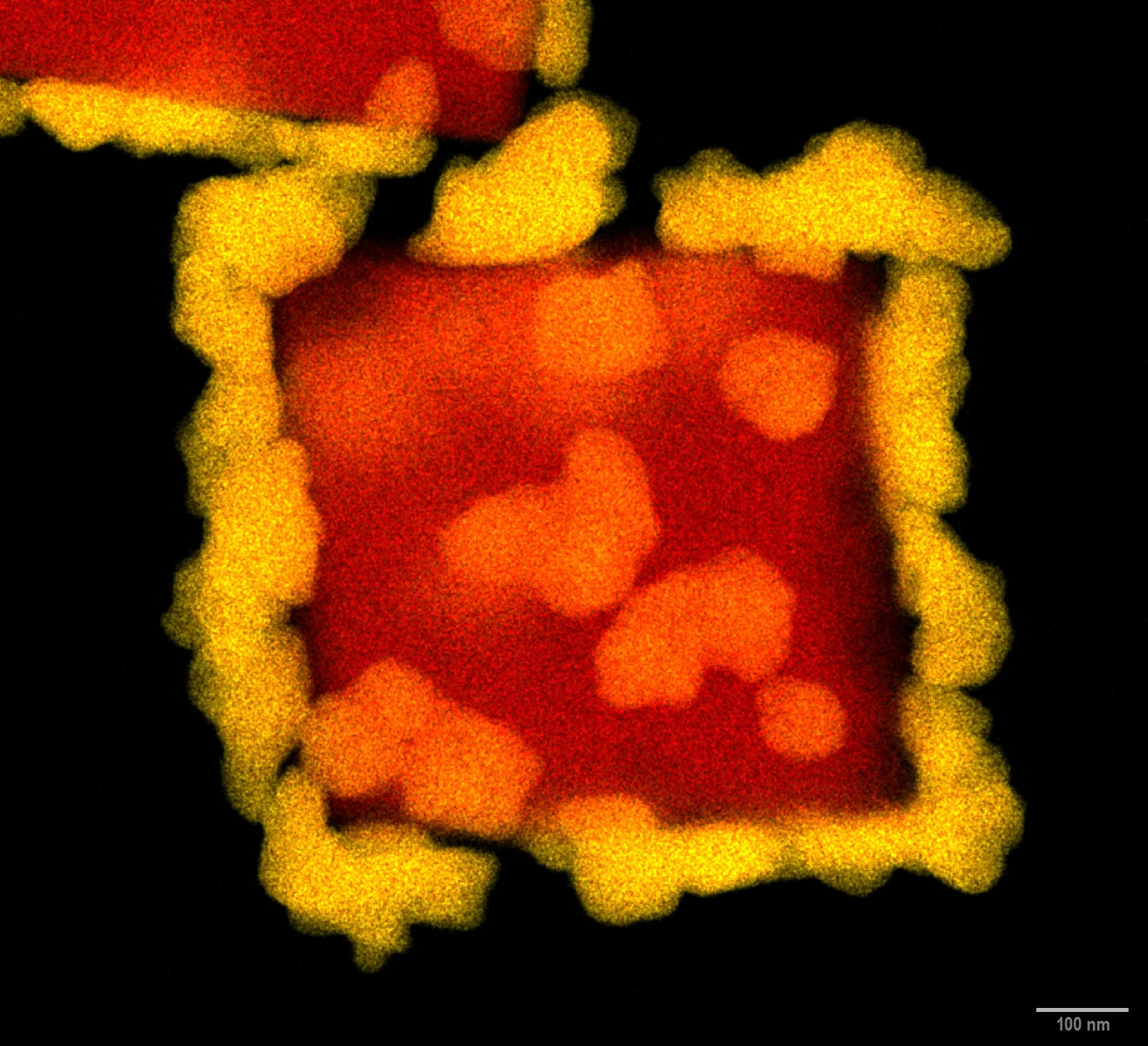
Copper cube decorated by gold nanoclusters, synthesized under the electron beam in basic environment. The image was acquired using energy dispersive X-ray spectroscopy mapping. False colors indicate the distributing of gold (yellow) and copper (red) in the investigated structure. (Source: IFJ PAN)
Beauty and mystery – it is hard to imagine a more alluring combination, and this is precisely what we encounter when we observe phenomena in the nanoworld. But are we really sure that observations made using modern microscopic techniques do not influence what is happening there? The answers are provided by Cracow scientists. A comparison of images of copper oxide nanoparticles reacting with chloroauric acid, obtained using electron and photon beams, has yielded unexpected results.
We have become accustomed to the idea that events occurring in the world around us do not depend on the manner in which we view them. But does a similar principle apply when, using modern microscopic techniques that utilize electron or photon beams, we attempt to observe the course of chemical reactions in the world of objects measuring in the nano- and micrometer range?
“We stumbled upon the clue that led to our discovery whilst investigating the galvanic exchange reaction
between copper oxide nanocubes and chloroauric acid. We examined this phenomenon in
a rather unusual way, using two sophisticated microscopic techniques: LC-TEM, or liquid-cell
transmission electron microscopy, and EL-STXM, or liquid-cell electrochemical scanning transmission
X-ray microscopy. In other words, at times we observed the process using electrons, and at
other times using photons in the X-ray energy range – and although in both cases we were observing
the same substances reacting with one another, we saw something different,
” explains Prof.
Magdalena Parlinska from the Institute of Nuclear Physics, Polish Academy of Sciences (IFJ PAN)
in Cracow, one of the lead authors of an article published in the prestigious scientific journal Nano
Micro Small.
In transmission electron microscopy (TEM), an electron beam in vacuum passes through a sample which is no more than a few dozen nanometers thick. Unfortunately, the vacuum inside the microscope prevents the examination of samples containing water or suspended in liquid. This problem is solved by holders featuring a liquid cell (LC) comprising two chips with windows fitted with two thin silicon nitride membranes, through which the imaging electron beam passes. In the experiment carried out by physicists in Cracow, the nanocubes were placed on a membrane, and a solution of chloroauric acid was delivered to the liquid cell via capillaries designed to supply fluids to the cell.
Scanning transmission X-ray microscopy (STXM) operates on a similar principle to TEM, but instead of electrons, it uses a focused beam of soft X-rays, i.e. photons. Given that the energies of the photons are significantly higher here than in the optical range, STXM also allows for high image magnification, albeit slightly lower than that achieved with electron microscopy. The technique does, however, have a major advantage: by scanning the sample with photon beams of appropriately selected energies, it is possible to obtain information about the material’s chemical composition and even its degree of oxidation at a specific point in the image. The source of X-ray radiation for STXM microscopy in the presented studies was the SOLARIS synchrotron, the main facility of the National Centre for Synchrotron Radiation at the Jagiellonian University in Cracow.
“In the reaction of copper oxide nanocubes with a solution of chloroauric acid that we are investigating,
the cubes themselves act as templates for the gold structures that are to be formed,
” explains Dr. Joanna Depciuch-Czarny (IFJ PAN), co-author of the discovery in question. “When we observed
the reaction using a photon beam, we ultimately saw a material composed of gold-rich, hollow
nanoboxes. But when we looked at the samples using an electron beam, the gold was simply
deposited on the walls of the copper nanocubes, and the cubes showed no sign of disappearing.
”
Why did a reaction that appeared to be exactly the same result in the formation of hollow gold nanoboxes (a material with a large surface area, potentially ideal for use as a catalyst, for example) in one case, whilst in the other the gold merely coated the copper nanocubes with a thin layer? The culprit turned out to be the manner, in which the researchers observed the reacting cubes.
Both the electron beam and the photon beam react with water. The reactive chemical species formed as a result of irradiation alter the local environment, favoring either the acceptance of electrons (reduction) or their release (oxidation); they also affect the activity of hydrogen ions – and thus the acidity or alkalinity of the solution, as measured by the widely used pH scale. It is known from literature on the subject that an electron beam creates a locally more acidic environment, which inhibits the oxidation and etching of copper oxide, whilst simultaneously promoting the reduction of gold ions and their deposition. In contrast, a photon beam does not cause such a drop in the solution’s pH as in the case of electrons, which aids both the deposition of gold and the oxidation/ dissolution of copper oxide, ultimately leading to the formation of hollow gold nanoboxes.
The experimental results were confirmed using numerical models describing ion transport, surface reactions and the radiolysis of water, carried out using the computing power of the Cyfronet AGH Academic Computing Centre.
“This article addresses fundamental issues: we have demonstrated that the beam used for microscopic
imaging of successive stages of chemical reactions need not be a passive observer. On the
contrary, it can become one of the actors on the chemical stage, capable of actively influencing the
course of events. We are already working on further publications, using materials containing other
elements and presenting the issue from a more applied perspective,
” concludes Prof. Parlinska.
The research presented here was funded by the Polish National Science Centre.
[PDF]
Contact:
Prof. Magdalena Parlińska
Institute of Nuclear Physics, Polish Academy of Science
tel.: +48 12 662 8160
email: magdalena.parlinska@ifj.edu.pl
Scientific papers:
„Electrons Make Shells, Photons Make Boxes: Unmasking Radiation Chemistry in Liquid-Phase Imaging”
B. Alavi, J. Depciuch, M. S. Shakeri, K. Sobczak, K. Matlak, M. Parlińska
Small 2026, e75209
DOI: 10.1002/smll.75209
Images:
Copper cube decorated by gold nanoclusters, synthesized under the electron beam in basic environment. The image was acquired using energy dispersive X-ray spectroscopy mapping. False colors indicate the distributing of gold (yellow) and copper (red) in the investigated structure. (Source: IFJ PAN)
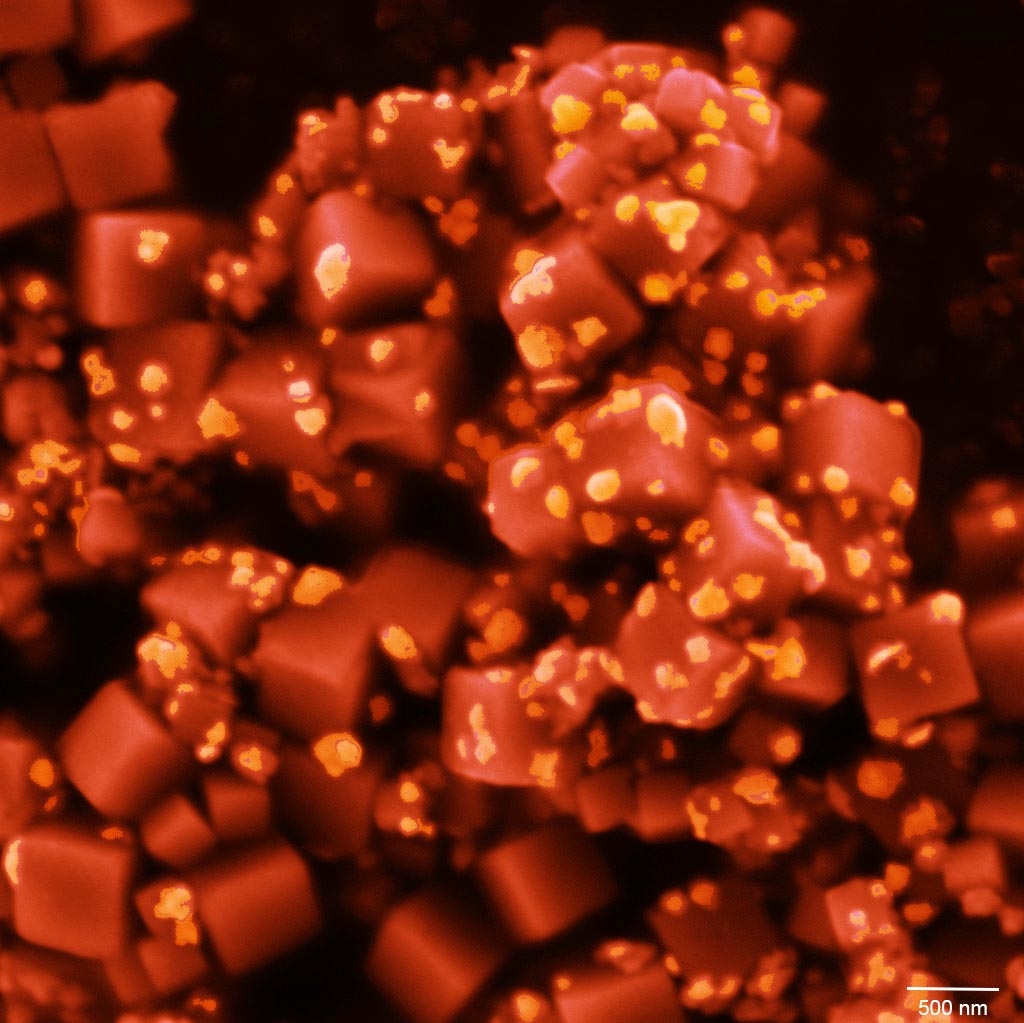
Gold nanoparticle-coated copper cubes. Artificially colored scanning electron microscope (SEM) image. (Source: IFJ PAN)