X-ray lasers: Why does brighter mean darker?
18 October 2023
EurekAlert!: [https://www.eurekalert.org/news-releases/1005250]

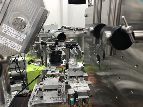
The SACLA free-electron laser facility, where the experiment on the diffraction of ultra-short X-ray pulses on crystalline silicon samples was carried out. (Source: SACLA)
When we illuminate something, we usually expect that the brighter the source we use, the brighter the resulting image will be. This rule also works for ultra-short pulses of laser light – but only up to a certain intensity. The answer to the question why an X-ray diffraction image 'darkens' at very high X-ray intensities does not only deepen fundamental understanding of the light-matter interaction, but also offers a unique perspective for the production of laser pulses that have significantly shorter pulse duration than those currently available.
The more light, the brighter? This observation might sound trivial, were it not for the fact that... it is not always true! When silicon crystals are illuminated with ultrafast laser pulses of X-ray light, the resulting diffraction images are indeed initially brighter the more photons fall on the sample, i.e., the higher the beam intensity. Recently, however, a counterintuitive effect has been observed: when the intensity of the X-ray beam starts to exceed a certain critical value, the diffraction images unexpectedly weaken. This puzzling phenomenon has just been explained, thanks to the efforts of the experimental and theoretical physicists from Japanese, Polish and German research institutions, including the RIKEN SPring-8 Centre in Hyogo, the Institute of Nuclear Physics of the Polish Academy of Sciences (IFJ PAN) in Cracow and the Center for Free-Electron Laser Science (CFEL) at the DESY laboratory in Hamburg.
X-ray free-electron lasers (XFELs) generate very powerful X-ray pulses with durations of femtoseconds, i.e., quadrillionths of a second. Machines of this type, currently operating at only a few locations in the world, are used, amongst others, to analyse structure of matter by means of X-ray diffraction. With this technique, a sample is illuminated by an X-ray pulse and the diffracted radiation is recorded. The obtained diffraction image is then used in order to reconstruct the original crystal structure of the material under examination.
“Intuition tells us that the more photons we have, the clearer the diffraction image of the sample should be. This is indeed the case, but only up to a certain X-ray intensity, of the order of tens trillions of watts per square centimetre. When this value is exceeded – and we have been only recently capable of doing this – the diffraction signal suddenly starts to weaken. Our research is the first attempt to explain this unexpected effect
”, says Prof. Beata Ziaja-Motyka (IFJ PAN, DESY), who deals with theoretical modelling and computer simulations of phenomena related to the interaction of ultrafast X-ray pulses with matter.
Theoretical research undertaken to explain the results of the experiment with XFEL laser firing on crystalline silicon samples at Japan's XFEL facility, called SACLA, Hyogo, has been supported by computer simulations. The following explanation of the phenomenon observed emerged from the researchers' work.
“When an avalanche of high-energy photons hits a material, there is a massive knockout of electrons from various atomic shells, resulting in a rapid ionisation of atoms in the material. Last year, our group showed that the first movements of ionised atoms in the crystal lattice, initiating the process of structural self-destruction of the sample, occured with a delay of approximately 20 femtoseconds after the light pulse hit the sample. We are now convinced that the reason for the recently observed weakening of the diffraction signal is due to phenomena occurring earlier, in the first six femtoseconds of the interaction
”, says Dr. Ichiro Inoue from the RIKEN SPring-8 Centre, responsible for the experimental study.
During the initial phase of X-ray-matter interaction, incoming high-energy photons rapidly excite not only 'surface' (valence) electrons from atoms, but also the electrons occupying deep atomic shells, located close to the atomic nucleus. It turns out that the presence of deep shell holes in atoms strongly reduce their atomic scattering factors, i.e., the quantities determining the intensity of the observed diffraction signal.
“Our research shows that before any structural damage to the material occurs and the sample disintegrates, first a rapid electronic damage occurs. As a result, the final part of the pulse practically no longer ionises the material, because further excitation of electrons by X-ray photons is no longer energetically possible
”, Prof. Ziaja-Motyka specifies.
At first glance, the observed effect appears to be just unfavourable, as it results in a decreased brightness of the diffraction images recorded. However, it seems that one can very well exploit this finding. The observation that different atoms respond differently to ultrafast X-ray pulses may help to more accurately reconstruct three-dimensional complex atomic structures from the recorded diffraction images.
Another area of potential application has to do with the production of laser pulses with extremely short pulse durations. Since the material through which the high-intensity X-ray pulse passes 'cuts off' a significant part of the already ultra-short pulse, it can be deliberately used as 'scissors' to generate pulses that are effectively shorter than those produced so far. If successful, this could stimulate another breakthrough in imaging of quantum world.
The research presented here was co-financed by the Institute of Nuclear Physics of the Polish Academy of Sciences.
[PDF]
Contact:
Prof. Beata Ziaja-Motyka
Institute of Nuclear Physics, Polish Academy of Sciences; Center for Free-Electron Laser Science CFEL, Deutsches Elektronen-Synchrotron DESY
tel.: +49 40 8998 6303
email: ziaja@mail.desy.de
Scientific papers:
„Femtosecond reduction of atomic scattering factors triggered by intense x-ray pulse”
I. Inoue, J. Yamada, K. J. Kapcia, M. Stransky, V. Tkachenko, Z. Jurek, T. Inoue, T. Osaka, Y. Inubushi, A. Ito, Y. Tanaka, S. Matsuyama, K. Yamauchi, M. Yabashi, B. Ziaja;
Physical Review Letters, 131, 16, 2023;
DOI: https://doi.org/10.1103/PhysRevLett.131.163201

